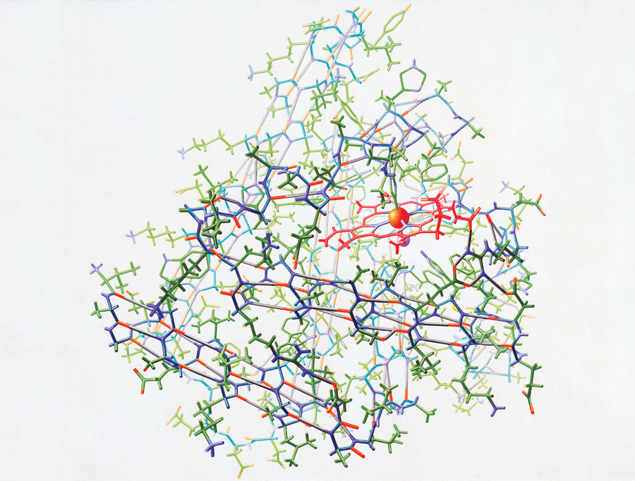
“Crystal structure of myoglobin (1961)” from the Irving Geis Collection. Rights owned and administered by the Howard Hughes Medical Institute. Reproduction by permission only.
The image above was a painting of myoglobin, the first protein structure solved by X-ray crystallography. The painting was created by Irving Geis for a Scientific American article “The Three Dimensional Structure of a Protein Molecule” by John Kendrew, published in December l961. John Kendrew and his colleagues solved the myoglobin structure in l958.

Irving Geis (1908-1997). Photo: Sandy Geis.
Nowadays, one can easily create an image of a protein structure with the aid of a computer and molecular visualization software. In 1961, however, everything had to be done by hand. Creating an image of a protein structure required not only outstanding artistic skills of visualizing complicated 3D structures, but also extraordinary patience. Originally trained as an architect at Georgia Institute of Technology and receiving a Bachelor of Fine Arts from University of Pennsylvania, Geis had all the skills and knowledge to visualize the 3D structures of proteins.
Geis created this painting by first photographing the physical models and then by creating voluminous sketches and studies before painting the finished version [1]. A lot of refinements were made during the sketch step based on the feedback from John Kendrew, as shown by the image below. The final painting took 6 months to complete [2].

“A draft image of the 1961 myoglobin” from the Irving Geis Collection. Comments on the image were made by Irving Geis and John Kendrew. Rights owned and administered by the Howard Hughes Medical Institute. Reproduction by permission only.
I tried to mimic Geis’ myoglobin image by using UCSF Chimera and Maxon Cinema 4D. It took me quite some time but the result is far inferior to the original masterpiece. The heme group seems OK, but the alpha helices are hard to recognize even though I used the same color scheme as Geis’ painting, depth cueing effect, and some lighting techniques in Cinema 4D. In addition, I couldn’t add hydrogen atoms except those in hydrogen bonds in my version, as the model would become overwhelmingly complicated to be meaningful at all. On the other hand, Geis’s painting clearly visualizes the main structural features (the heme group and alpha helices) while giving an overall sense of the structural complexity of the myoglobin protein.
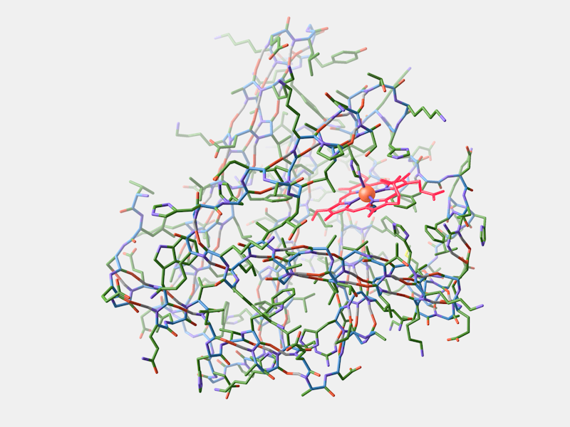
My attempt to mimic Geis’ painting using UCSF Chimera and Maxon Cinema 4D (PDB ID: 1MBN). The alpha helices are difficult to recognize because they overlap with the atoms in the back.
To achieve this, Geis used a process he called “selected lying” [2-4], in which he made small adjustments to avoid structural overlapping. He might distort the protein a little bit here and there, or he might use slightly different viewing angles or perspectives for different parts of the protein. In the end, this process resulted in a structure that was not so different from the real structure, but much easier to understand on a flat paper. A computer, on the other hand, draws everything based on the given coordinates and the image it produces is usually not very comprehensible, especially for complicated protein structures.

“Crystal structure of lysozyme (1966)” from the Irving Geis Collection. Rights owned and administered by the Howard Hughes Medical Institute. Reproduction by permission only.
Besides his excellence in visualizing complicated 3D structures, what makes Geis’ paintings special is his belief that “his job was not to draw a protein exactly as it was, but to show how it worked”[3]. Often Geis would add additional layers of information on top of protein structures, making protein functions and mechanisms understandable. This is the reason why his protein paintings are still appreciated and used in some textbooks, even today.
Colors are very important to Geis’ painting. He carefully chose the proper colors to illustrate the inner workings of proteins. “Color is a language”, he said, “and as with any other language, one mustn’t babble!” [3]

“Cytochrome C (1989)” from the Irving Geis Collection. Rights owned and administered by the Howard Hughes Medical Institute. Reproduction by permission only.
Geis had worked with many scientists in his career. Among them, Dr. Richard Dickerson was his long time collaborator and friend. The two co-authored several books, including The Structure and Action of Proteins (1969), Hemoglobin (1983), and a chemistry textbook: Chemistry, Matter and the Universe (1976). The Structure and Action of Proteins became classic after published and inspired a generation of young biochemists.
Irving Geis was born in 1908 in New York City and died in 1997. He was 88 and lived in Manhattan.

“Irving Geis and his work-in-progress 1961 myoglobin painting” from the Irving Geis Collection. Rights owned and administered by the Howard Hughes Medical Institute. Reproduction by permission only.
Interview with Dr. Richard E. Dickerson about Mr. Geis
1. It’s rare that an artist shares the authorship with a text writer. However, you and Mr. Geis coauthored 3 books together [The Structure and Action of Proteins (1969), Chemistry, Matter and the Universe (1976), Hemoglobin (1983)]. As you wrote, “I could describe what needed to be illustrated about protein structure, and Irv would come up with clever and original graphic methods of putting the point across. It was never clear whether Irv illustrated my books, or I wrote Irv’s captions.”[3] Could you please share with us how Mr. Geis was able to get such a deep understanding of proteins?
It is my understanding that he acquired his background knowledge slowly by illustrating for magazines such as Scientific American. Indeed, it was his illustrations for John Kendrew’s myoglobin structure that first caused us to meet in New York in 1963. He was a very intelligent man, and simply read a lot about scientific matters in order to make his diagrams more meaningful.
2. In you books, I am really amazed that Mr. Geis was able to depict sophisticated protein structures and functions with only black/gray and another color. The images are not only easy to understand but also elegant. Could you please comment on this?
Irving Geis was a true artist. A skillful artist can convey information that a simple draftsman cannot. His use of color and shading for emphasis was masterful.
3. As you wrote, Mr. Geis used a process he called “Selective Lying” to tweak the protein representation if “some key aspect of protein structure was eclipsed and out of sight”.[3] The end result is a protein structure and its molecular mechanism easy to understand. Personally, I am firm believer of this approach. But others might think we should not alter the structure at all. What is your opinion on this?
The purpose of Irv’s scientific drawings was to convey information about how the molecules work, and not simply to illustrate the fine-structure details of the molecules. For the latter, one should turn to the Protein Data Bank (PDB) or other such archives. If a certain amount of artistic licence would aid the reader in understanding how the molecule worked, then such behaviour is not only legitimate; it is constructive.
4. As our knowledge of protein structure and function grow rapidly, computer animation becomes a common media to depict the dynamic protein behaviours in the cell. Compared with traditional protein illustrations, what are the pros and cons of computer animations? What would be the basic requirements for artists/scientists creating these animations?
I have no strong opinions on this. If computer animation is available to a research worker or a student, then it obviously could assist him in understanding the macromolecule.
5. You mentioned in an article[5] that you regretted you were not able to write Atlas of Protein Structure with Mr. Geis at a time when there were only 8 protein structures. Nowadays, we have nearly 100 thousands protein structures in the Protein Data Bank. Do you think it is still necessary write a new Atlas to give students and hopefully the general public a feel of the richness and magic of protein structures? If you do, how many proteins should be included in this challenging book?
I think that the days of an atlas that contains every known structure are over. No one can write a book that discusses 100,000 different protein structures. The PDB is a resource that allows people interested in a certain class of protein to learn how many such proteins have been solved and what each of them looks like. It is true that one could write a general book on protein structure/function relationships, and indeed some have done so. Such authors would select those of the 100,000 different structures that pertained to the matter at hand, or a set of structures that gave a general impression of protein structure. But to discuss all 100,000 structures individually: No.
6. Finally, what is your favorite illustration(s) of Mr. Geis and why?
I simply could not come up with one particular “favorite” illustration; Irv has done so much and done it so well. I like his haemoglobin/myoglobin and his DNA illustrations, because these concern subjects in which I had a scientific relationship. But someone else would undoubtedly give you a different list.
Acknowledgement
I would like to thank Sandy Geis for reviewing this article and generously providing many reference materials about Mr. Geis’s works. I would also like to thank Howard Hughes Medical Institute for granting permissions to use the above images from the Irving Geis Collection. Finally, I am grateful that Dr. Dickerson took the time to answer my questions.
Rreferences
[1] S. de Chadarevian, Models and the Making of Molecular Biology, in Models: The Third Demension of Science (eds. S. de Chadarevian and N. Hopwood) 339-368 (Stanford Univeristy Press, Stanford, California, USA, 2004)
[2] B. P. Gaber and D. S. Goodsell, Irving Geis: Dean of Molecular Illustration. Journal of Molecular Graphics and Modeling 15, 57-59 (1997)
[3] R. E. Dickerson, Irving Geis, Molecular artist, 1908-1997. Protein Science 6, 2483-2484 (1997)
[4] D. S. Goodsell and G. T. Johnson, Filling in the Gaps: Artistic License in Education and Outreach. PLoS Biology 5, 2759-2762 (2007)
[5] R. E. Dickerson, Obituary: Irving Geis, 1908-1997. Structure 5, 1247-1249 (1997).